
Hardware Solutions
Applications
 Part of the Oxford Instruments Group
Neuroscience
The naturally circulating hormone estradiol is required for female sexual behavior in rodents, but thus far there hasn’t been evidence that this hormone affects the structure of motivational circuitry. Researchers in Robert L. Meisel’s laboratory at the University of Minnesota recently analyzed dendritic spines with Imaris software and found that estradiol does significantly alter the neuronal architecture of a key reward area in the brain of female hamsters.
“Our laboratory utilizes female sexual behavior as a model of naturally motivated behavior in effort to study the neurobiology of motivation,” said Nancy A. Staffend, a member of the research team. “The overarching goal of the laboratory is to delineate the neurobiological consequences of naturally motivated behavior on the motivational circuitry to parse out the differences between natural behavior and pharmacological activation through drugs of abuse.”
Using Imaris’ FilamentTracer the researchers looked for structural changes in response to estradiol in medium spiny neurons of the nucleus accumbens, a vital reward area in the brain. They used DiI to label neurons from the nucleus accumbens of estradiol-treated female hamsters and then imaged the neurons using confocal microscopy. With the FilamentTracer’s AutoDepth feature they manually traced 3-D images, assigning the dendrite and spines in the XY plane. The center function let them automatically center the manually traced dendritic branches, accurately aligning the drawing with the dendrite and spines. They then used the software’s diameter function to create an accurate 3-D model of the dendritic branches and spines.
The group measured the dendritic length and number of spines in the 3-D rendering and used this information to calculate the spine density for every 10 microns of dendrite. In addition, they used the Imaris XTension, Classify Spines, to categorize spines as stubby, filopodial, long/thin, or mushroom-shaped to determine differences in spine morphology.
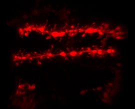
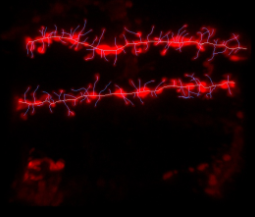
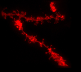
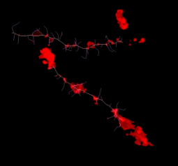
Figure 1 - Two dendritic segments are shown in the absence (top) and presence (bottom) of the hormone estradiol. The left images show just the dendritic segments and the right images show dendritic segments with the manually traced "skeleton" of the dendrite and spines visualized with Imaris FilamentTracer. After estradiol treatment, the reduction in dendritic spine density of medium spiny neurons within the core of the nucleus accumbens is evident.
Staffend said that the primary reason their laboratory purchased Imaris 3D and 4D image analysis software is its automation capabilities. “The 3-D renderings allow us to not only attain information regarding spine density but also to perform high-throughput analysis of dendritic spine morphology,” she said. “Manual morphological measurements require a tremendous amount of time, and although we do use manual tracing to establish our initial dendritic “skeleton,” the automation that follows makes our analysis stream lined and efficient.”
Their analysis showed that estradiol significantly reduces dendritc spine density, specifically within the core region of the nucleus accumbens. This suggests that estradiol may impact the motivational state of the female on a structural level. “We are currently taking advantage of additional information that the Imaris software generates including Scholl analysis, dendrite diameter, and spine terminal point diameter in an effort to further delineate structural changes which could potentially suggest a change in function,” Staffend said.
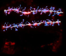
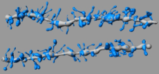
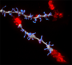
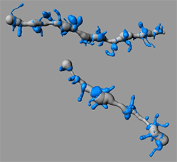
Figure 2 - The top images show dendritic segments in the absence of estradiol, and the bottom images are with estradiol present. Dendrite segments with the Imaris-generated 3D rendering of the dendrite and spines are on the left. The image analysis was performed using the 3D renderings alone (right).
Similar changes in neural structure occur in response to repeated exposure to naturally motivated behaviors and repeated exposure to drugs of abuse. To this end, the researchers are evaluating structural changes that occur in female hamsters following repeated exposure to sexual behavior or aggression.
Research Paper: Estradiol reduces dendritic spine density in the ventral striatum of female Syrian hamsters, Brain Structure and Function, Vol. 215, Numbers 3-4, pp. 187-194, DOI: 10.1007/s00429-010-0284-7.
© Oxford Instruments 2024
