
Hardware Solutions
Applications
 Part of the Oxford Instruments Group
Cell Biology
Invariant natural killer T cells are a subset of T lymphocytes involved in the host defense against microbial infection. These T cells recognize glycolipids bound to the surface molecule CD1d, which is expressed by antigen-presenting cells. Scientists would like to understand the dynamics of how and where these T cells encounter antigen and become activated in vivo.
To find out more about the interactions of invariant natural killer T cells with antigen, Dr. Patricia Barral from the Cancer Research UK and her colleagues used multi-photon microscopy imaging. “To mount a protective response to infection, lymphocytes rapidly alter their behavior after antigen encounter in terms of motility, localization and interaction with other cells,” said Barral. “Multi-photon microscopy allows the study of the dynamic behavior of lymphocytes within their natural environment in response to infection.
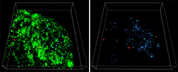
Figure 1: Tracking of natural killer T cells in lymph node using Imaris. Invariant natural killer T cells (red) were adoptively transferred into wild-type recipients with wild-type B cells (blue). Lipid antigen (green) was injected 16 h after adoptive transfer. Invariant natural killer T cells are confined in the lymph node periphery in response to lipid antigen and get arrested around antigen-rich areas. A 3-D reconstruction of a lymph node region is shown with the tracks of natural killer T cells (pale red) and B cells (blue) during a 20-minute imaging period.Images courtesy of Patricia Barral, Cancer Research UK.
The researchers used multi-photon microscopy to image through the capsule of a lymph node, acquiring stacks of 11 to 20 square xy planes spanning 508 × 508 μm with 5-μm z spacing over 20 to 30 min. They transformed the image stacks into volume-rendered 4-D movies and analyzed them using Imaris. The software was used to automatically track cells and to calculate average cell speed, instantaneous speed, the confinement index, and displacement.
Three-dimensional reconstructions of lymph nodes made using Imaris allowed the researchers to determine the location of natural killer T cells relative to CD169+ macrophages from the lymph node periphery. They found that in resting conditions, the invariant natural killer T cells resided mainly in the paracortical region of the lymph nodes, while searching for specific antigen. Within minutes of antigen administration, the antigens arrived at the lymph nodes and were retained by CD169+ macrophages in the lymph node periphery. This induced arrest of the invariant natural killer T cells in this region as soon as 2 hours after antigen administration in a CD1d-dependent manner.
“Furthermore we found that CD169+ macrophages retain lipid antigen and engage in long-lasting interactions with invariant natural killer T cells, which results in very fast invariant natural killer T cell activation and cytokine secretion,” Barral said. “Thus, we have identified CD169+ macrophages as bona fide antigen presenting cells controlling early invariant natural killer T cell activation and favoring fast initiation of immune responses.”
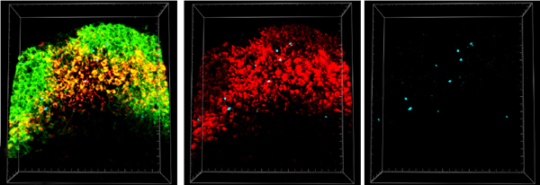
Figure 2: Arrest of invariant natural killer T cells in CD169+ macrophages. The killer T cells (blue) were adoptively transferred into wild-type recipients that were afterwards immunized with lipid antigen (red). CD169+ macrophages were visualized by administration of fluorescently labeled anti-mouse CD169 antibody. This 3-D reconstruction of a lymph node section (100 mm thick) shows that invariant natural killer T cells were visualized in close contact with antigen loaded-CD169+ macrophages. Images courtesy of Patricia Barral, Cancer Research UK.
Research Paper: P. Barral, P. Polzella, A. Bruckbauer, N. van Rooijen, G. S. Besra, V. Cerundolo & F. D. Batista CD169+ macrophages present lipid antigens to mediate early activation of iNKT cells in lymph nodes. Nature J. of Immunology 11(4),303-312 (2010)
© Oxford Instruments 2024
