
Hardware Solutions
Applications
 Part of the Oxford Instruments Group
Stem Cell
Sean J. Morrison and colleagues at the University of Texas Southwestern Medical Center are using Imaris to study the stem cells that give rise to the various types of blood cells found in humans. Understanding how these cells, known as hematopoietic stem cells, interact with their environment could inform new methods that treat blood system diseases such as anemia or cancer by manipulating the stem cell environment.
Although hematopoietic stem cells are one of the most well characterized types of adult stem cells, the specific location where they reside within the bone marrow has remained controversial. “Combining advanced deep confocal imaging of bone marrow and the powerful analysis tools of Imaris allowed us to comprehensively determine the distance, in three dimensional space, of thousands of hematopoietic stem cells relative to different blood vessel types, perivascular stromal cells, and bone,” said Malea Murphy, a member of the research team.
The researchers used a clearing technique that allowed deep confocal imaging within a large segment of bone marrow. After acquiring z-stacked optical sections up to 600 microns deep into the bone marrow, they used the Imaris to render 3D reconstructions. They then turned to the Imaris Spots function to mark the size and location of each stem cell and the Imaris Surfaces function to create digital Surfaces on the three types of blood vessels (arterioles, the connecting capillaries, and venous sinusoids), perivascular stromal cells, and bone.
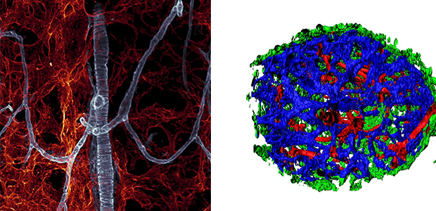
The left image shows sinusoidal blood vessels in red and arteriolar blood vessels in blue. For the right image, the researchers used the Imaris Surfaces segmentation to create digital Surfaces on the three types of blood vessels (red=arteriolar , blue=sinusoidal, green=connecting capillaries).
Calculating distance in large datasets
Imaris allowed the researchers to easily work with the large stacks of tiled confocal imaging data (H:3mm, W:1mm, D:0.5mm), in which they visualized the stem cells in 3D space. They used the Imaris Distance Transform Matlab® XTension function to calculate the distance in 3D space from each Surface to every stem cell Spot and conducted volume calculations using a Matlab®-based Imaris XTension.
“The flexibility of the Imaris Surfaces detection allowed us to use a single fluorescent marker for all blood vessels and then use morphology and location to create independent Surfaces of the different vessel types,” Murphy explained. “The Distance Transformation XTension function is much better than relying on vector distances annotated by eye. We easily exported the minimum, maximum, and center distances for each stem cell as an Excel file for each sample, which really facilitated analysis of these large data sets.”
Overall, the analysis revealed that hematopoietic stem cells were more commonly found in the central marrow rather than near bone surfaces and that approximately 85 percent of the stem cells were within 10 mm of a sinusoidal blood vessel. Although some scientists had thought that dividing and non-dividing hematopoietic stem cells were located in different areas of the bone marrow, the new study showed that both dividing and non-dividing hematopoietic stem cells were typically found some distance away from arterioles, transition zone vessels, and bone surfaces, thus residing mainly in the perisinusoidal niches throughout bone marrow.
© Oxford Instruments 2024
